Scientific Program

Conference Series Ltd invites all the participants across the globe to attend 3rd European Otolaryngology-ENT Surgery Conference London, UK.

Day 2 :

Keynote Forum

Alessandro Bucci

Hospital of Senigallia, Italy

Keynote: An updated review of DISE
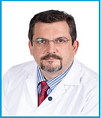
Conference Series ENT 2018 International Conference Keynote Speaker Alessandro Bucci photo
Biography:

Alessandro Bucci pursued his PhD (Rhinology and Rhino-Allergology) in 2006 at UCSC (Catholic University), Rome (Italy). He completed his Residency in Otolaryngology-Head and Neck Surgery at UCSC - Gemelli Hospital in Rome. His fellowship credentials include: Fellowship in Otolaryngology in Spain (University Hospital, Cadiz); Fellowship trained in Facial Plastic Surgery (AMC) and OSAS (Sint Lucas Andreas Hospital) in Amsterdam, The Netherlands; Facial Plastic Surgery (Calixto Garcia
University Hospital), Havana, Cuba. He is currently the Head of Sleep Apnea Center and Rhinology/Rhino-Allergology Center in the Otolaryngology Department of ASUR Marche AV2, Senigallia, Italy. He is an International Faculty Member of the XXXV Pan-American Congress of Otorhinolaryngology 2016, Cuba. He has been the past
Director of the 1st International Conference on Rhinology and Rhino-Allergology; 5th Bulgarian Italian Rhinology Meeting, 2016 Senigallia (Italy). His credentials include: University Professor at the Marche Polytechnic University, Ancona, Italy; International Faculty Member of the VI Bulgarian Italian Meeting of Rhinology, Medical School: Catholic University (UCSC) of Rome; He served as Reserve Medical Officer of the Italian Navy. His research focuses on Rhinology/Rhino Allergology, Sleep Apnea/OSAS. His main interest include: humanitarian and international outreach. He served as: Vice-President of the National Association of Tracheotomized patients- ONLUS ( nonprofit organization of social utility); Member of the ERS (European Rhinologic Society).

Abstract:

Obstructive sleep apnea (OSA) is characterized by intermittent, repeated upper airway narrowing or obstruction occurring during sleep. Most authors agree that rigorous patient selection is logical and mandatory. Successful surgical treatment of OSA is based on the accurate identification of the pattern of airway obstruction and targeted, effective treatment. Nevertheless, surgical results in OSA vary greatly, whatever the surgical technique or site treated. For many years, the primary surgical treatment for obstructive sleep apnea was soft palate surgery, and this worked well for patients with blockage of breathing in the palate region alone. Unfortunately, many patients also appear to have blockage of breathing in the tongue region, and multiple procedures have been developed to address this in the hope of improving surgical outcomes. DISE (Drug induced sleep endoscopy) was introduced almost two decades ago and used extensively since the 2000s. Nevertheless, there are still limited data about the real role of sleep endoscopy in obstructive sleep apnea syndrome. DISE offers a unique structure-based assessment of the airway, compared to other commonly-used evaluation techniques. It mimics sleep in order to observe the upper airway on flexible endoscopy. Because surgical procedures are ultimately directed at specific structures, DISE may improve procedure selection and outcomes. DISE involves a certain number of limitations: natural sleep is not precisely reproduced, the anesthesia protocol is not standardized, and there is no gold-standard validation. Classification is imperfect, indications remain to be standardized. Despite the lack of standardization in clinical examinations, the type of drugs used for sedation and the classification system used, the results obtained till date favor the inclusion of DISE in the investigation of obstruction sites in patients with OSA.

Keynote Forum

George J. Bitar

Bitar Cosmetic Surgery Institute, USA

Keynote: Ethnic Rhinoplasties: A 16 year experience
Conference Series ENT 2018 International Conference Keynote Speaker George J. Bitar photo
Biography:

George J. Bitar, MD, FACS is an award-winning, the medical director, fellowship-trained, board certified plastic surgeon and has performed over 5,000 cosmetic facial and body procedures. Dr. Bitar obtained his medical degree from the George Washington University School of Medicine in Washington, DC, where he now serves as an Assistant Clinical Professor. He then finished a general surgery residency at the Albert Einstein Medical Center in Philadelphia. His plastic surgery residency was completed at the University of Virginia Plastic Surgery Program, which provided him with a strong foundation in reconstructive surgery, for people injured in accidents or who have facial deformities or severe burns, and in hand surgery and microsurgery.

Abstract:

Introduction: Performing ethnic rhinoplasties successfully requires many skills. Technical expertise is must. A plastic surgeon should have the skill-set to perform a rhinoplasty and accept to perform such surgeries commensurate with his or her level of expertise, resulting in naturally looking results. Various ethnic rhinoplasties include Caucasian nose (European white), Hispanic nose, Middle Eastern nose (Arabic, Turkish, North African, and Persian Descent), Asian Nose (Chinese, Japanese, Filipino, Korean, and Indonesian) and African nose.
Methods: Non-surgical algorithm: Nonsurgical or filler rhinoplasty is becoming more popular due to safe and readily available fillers. Injectables are commonly done on all nose types with calcium based fillers for primary noses or when more volume needs to be achieved, and hyaluronic acid fillers for primary noses that need contour changes or secondary noses with concern for blood supply. Surgical algorithm: A nose should be thought of as a pyramid and should look good from all angles after a rhinoplasty is completed. My algorithm for rhinoplasty will be discussed.
Conclusion: Ethnic rhinoplasty is a precise and demanding operation due to many factors discussed in this talk. The upside is that
if all the considerations are addressed, then our patients will be very happy and have noses that match their identity and sense of
beauty.

Keynote Forum

Santosh Kumar Swain

Siksha O Anusandhan University, India

Keynote: Primary laryngeal tuberculosis: Experiences at a tertiary care teaching hospital of India

Time : 10:30-11:00

Conference Series ENT 2018 International Conference Keynote Speaker Santosh Kumar Swain photo
Biography:

Santosh Kumar Swain pursued MS in Otorhinolaryngology from VSS Institute of Medical Sciences and Research, India; completed Senior Residency from Christian Medical College, Vellore, India; passed Diplomate of National Board (DNB) in Otorhinolaryngology. He is the Head of the Department of Otolaryngology and Head-Neck
Surgery at the Institute of Medical Sciences and SUM Hospital, Siksha O Anusandhan University, India. He is a Member of National Academy of Medical Sciences, New Delhi. Currently, he is also working as a mentor for the Department of Science and Research Government of India on the research project: “Development of molecular kit to minimize the ciprofloxacin and amoxyclav resistance strains from chronic suppurative otitis media.” He has published more than 70 research articles in peer reviewed journals. In 2017, he was awarded Medical Talent of the Year at New Delhi, India.

Abstract:

Primary laryngeal tuberculosis is a chronic bacterial infection of the larynx by Mycobacterium tuberculosis without affecting the lungs. It is a rare type of extrapulmonary tuberculosis seen in clinical practice. This study aimed to evaluate the clinical presentation, diagnosis, and treatment of primary laryngeal tuberculosis at a tertiary care teaching hospital in eastern India. This is a retrospective study of 11 cases of primary laryngeal tuberculosis managed between December 2013 and January 2018. The detailed clinical presentations, investigations, and treatment of primary laryngeal tuberculosis of the patients were studied. Primary laryngeal tuberculosis is common in men with mean age of 38.63 years. Hoarseness of the voice is the most common symptom, and the most common site for primary laryngeal tuberculosis is the vocal fold with ulcerative lesion. Endoscopic examinations of the larynx in laryngeal tuberculosis are nonspecific and are to be confused with laryngeal cancer. Histopathological and bacteriological examinations are confirmatory tests for the diagnosis. After confirmation of the diagnosis, all patients had taken antitubercular therapy for 6 months, which gave excellent outcome. Delayed diagnosis or untreatable laryngeal tuberculosis will lead to high morbidity and mortality of the patient. Although primary laryngeal tuberculosis has nonspecific clinical presentations, it is very important to have a high index of suspiciousness to rule out tubercular lesion in the larynx as this disease is curable.

  • Head and Neck Surgery and Oncology| Oral and Maxillofacial surgery | Otolaryngology | Craniofacial surgery | Surgery For Nasal Disorders | Dentistry
Location: Bleroit 1
Speaker

Chair

Mark Shikowitz

The Feinstein Institute of Medical Research, USA

Speaker

Co-Chair

George J. Bitar

Bitar Cosmetic Surgery Institute, USA

Session Introduction

Naser Azmi Khayat

Arab American University, Palestine

Title: Diagnosis and treatment on temporomandibular disorders and Sleep apnea
Speaker
Biography:

Naser Azmi Khayat has been working as a lecturer in the Arab American university, Palestine. He did his B.Sc. in Dentistry from Baghdad University in Iraq in 1998 and M.Sc. in Orthodontics from AL-Mosul University in Iraq in 2000. He is also the Chairman of “Palestinian Academy for TMD & Dental Sleep Medicine” from 2018 and since 2014 until now he has been doing PhD researches in the relation between orthodontic problems and temporomandibular disorders at Tel Aviv University.

Abstract:

Temporomandibular disorder (TMDs) is a term for pain and dysfunction involving the masticatory muscles and the temporomandibular joints (TMJs) and is the most common orofacial pain condition. Prominent features are regional pain in the face and preauricular area, limitations in jaw movements and noise from the TMJs during jaw movements. TMD affects up to 15% of adults, with a peak incidence at 20 to 40 years of age. Chronic pain is the overwhelming reason for patients with TMD treatment. TMD can be associated with impaired general health, depression and other psychological disabilities and affect quality of life and well-being of the patient. The recently published diagnostic criteria for TMD (DC/TMD) have been found tobbe reliable and valid. These criteria include the most common types of TMDs including pain-related disorders, such as myofacial pain, headache attributed to TMD, arthralgia, as well as disorders associated with the TMJ, primarily disc displacements and degenerative disease. Co-morbid pain conditions such as headache, back and neck pain are common among TMD patients. Of this headache is most commonly reported and a significant overlap between headache and TMD-pain has been reported. Both conditions involve trigeminal neural processes, are characterized by pain in the head and/or face and pericranial tenderness. The primary utility of the DC/TMD diagnosis headache attributed to TMD is that it points to TMD treatment as a therapeutic approach for headache. Most patients with TMD improve with a combination of noninvasive therapies, including behavior therapy, pharmacotherapy, physical therapy and occlusal appliances. Obstructive sleep apnea (OSA) is a disorder in which recurrent closure of the upper airways occur during sleep. It is characterized by repetitive episodes of complete (apnea) or partial (hypopnea) upper airway obstruction occurring during sleep. Whereas apnea is complete cessation of airflow, hypopnea is characterized by a 70% reduction of airflow for ≥10 seconds or any reduction in airflow that is associated with either an
arousal from sleep or a ≥3% arterial oxygen desaturation. These frequent arousals are the primary cause of excessive daytime somnolence, are associated with impaired daytime cognitive function and are recognized as a cofactor in the etiology of road traffic accidents. Sleep apnea may also be associated with increased cardiovascular and increased cerebrovascular morbidity and mortality. An estimated 82% of men and 92% of women with moderate to severe sleep apnea remain undiagnosed. OSA treatment is chosen is based on patients clinical features. Its resolution may occur by losing weight, if the patient is obese, avoiding sleep in the supine position, if the OSA is position dependent, avoiding alcohol consumption during the evening and treating hypothyroidism if present. When these approaches are proved to be unsuccessful, other treatment should be chosen:Nasal continuous positive pressure (CPAP), surgical treatment or the application of oral appliances. Actually the role of a dentist is multi-level both in clinical evaluation and in treatment of OSA.

Speaker
Biography:

Robert J. Wood, has been appointed as the Associate Medical Director Craniofacial and Plastic Surgery, Gillette Children's Specialty Healthcare in 2014. He is also the Medical Director, Minnesota Centre for Craniofacial Services since 1999. His interests in the field are mostly with Craniofacial and Plastic Surgery.

Abstract:

Primary open repair of craniosynostosis has reported transfusion rates approaching 100%. We have developed a multimodal approach to blood conservation in an attempt to decrease rates of transfusion. Children aged one year or under undergoing primary repair of craniosynostosis from 10/1/13 to 1/15/16 received a multimodal approach to blood conservation including: pre-operative erythropoietin and oral ferrous sulfate, tumescent infiltration of the scalp incision, plasma blade incision and dissection, modest hemodilution and I V tranexamic acid. All children with an HgB less than 7g/dL or symptomatic of anemia were transfused. Surgical technique remained unchanged with classic fronto-orbital advancement and anterior cranial vault remodeling for anterior craniosynostosis, posterior calvarial remodeling for lambdoid and sagittal craniosynostosis including barrel stave type osteotomies, out fracture of the cranial base and interposition bone grafts. 96 children underwent repair with a mean age of 0.51 years. 74 (77%) did not require transfusion. Posterior repairs (n=55) averaged 6.5 mL/kg blood loss and 18% transfusion rate. Anterior repairs (n=41) averaged 9.1 mL/kg blood loss and 29% transfusion rate. Operative time averaged 55 minutes for posterior repairs and 82 minutes for anterior repairs. This is a significant advance in blood conservation.
Transfusion rates have dropped 82% for our posterior repairs. As experience is gained we expect further decreases in rates of
transfusion.

Speaker
Biography:

Yasser Mohamed Elsheikh is awarded Doctor Degree in Plastic, Oral & Maxillofacial Surgery from Alexandria University, Egypt in 2006. He holds a Master Degree (MSc) in General Surgery from Alexandria University, Egypt in 1997, followed by a Bachelor’s Degree (BSc) in E.N.T. from Alexandria University, Egypt in 1992. Currently, he is working as an Associate Professor for MENOUFIA UNIVERSITY. His international experience includes various programs, contributions and participation in different countries for diverse fields of study. His research interests as an Associate Professor reflect in his wide range of publications in various national and international journals. He is the Board Member of Member of Egyptian Society of Plastic and Reconstructive Surgery and serves as a member of Pan Arab Association and Egyptian Society of Oncology.

Abstract:

Surgery for saliva control has been available. A variety of operations have been tried, some being more successful than others. The surgery that is currently aims to redirect the saliva sitting at the front of the mouth to going back down the throat. There are 3 major pairs of glands in the mouth, the submandibular, sublingual and parotids. The submandibular and sublingual glands produce saliva through ducts in the front of the mouth just under the tongue. The submandibular produces most of the saliva in the mouth (about 65%) and it is watery. The sublingual produce a little saliva but it is thick and mucousy. The parotid glands produce saliva through ducts which open into the mouth near the second upper molar tooth. The saliva from the parotid is very helpful in making the food go down when we eat. The current study entails five patients complaining of sialorrhea and saliva drooling and one patient complain of postoperative atresia of both orifices of submandibular ducts and one patient has a suspicious swelling over the orifice of the left submandibular duct. Rerouting of submandibular duct was a very successful operation to solve their salivary problems.

Biography:

Muayed M. Radi Alkhafaji received his Medical Degree from University of Al- Mustansiriyah Medical School, (1986) and Board Certificate in ORL-HNS from Baghdad University, Iraq (1996) respectively. He is an ENT-Otolaryngologist in Doha, Qatar and is affiliated with multiple hospitals in the area, including Hamad General Hospital, Ambulatory Care Center, Alkhor Hospital and Weill Cornell Medical School-Qatar.

Abstract:

HFNC is a recently described technique proven to provide an adequate oxygenation & ventilation in patient undergoing various upper air way surgeries without the need of the endotracheal intubation or jet ventilation. The objective of this research is to evaluate the adequacy of HFNC technique as a sole method of providing adequate Oxygenation & ventilation for patients undergoing upper air way endoscopy (Micro laryngoscope, esophagoscope, Panendoscope) which are minor procedures need short duration of GA. Because the anesthetist & the surgeon sharing the same space there is high risk of interruption of ventilation, oxygenation, loss of airway in addition to inherent complication of the surgery. HFNC has the advantage of less interference during the procedure and gives large space for surgical manipulation and lastly decreases the intubation complication. This clinical trial will give additional impact of this treatment on ventilation (CO2 elimination) and oxygenation of the patient during prolongs apnea. The methodology involved a prospective, randomized interventional study using of computer program for randomization. The sample size for this study is 60 (30 in each group). Study is going on right now at (ACC) Ambulatory Care Center at Doha, Qatar and it will be completed within a year. The following criteria were taken into consideration: Inclusion criteria: adult population - both male and female - ASA 1 and 2 (American Society of Anesthesiologist) – for upper airway procedures. Exclusion criteria: age <18 years - BMI >35; for pregnant patient - procedure anticipation >45 min - total airway obstruction; for patient with sever nasal obstruction- parameters: age, gender, height, weight, BMI, HR, Bp, SPO2, TcPO2, ETCO2, TcPCO2 and BIS. Trans nasal Tracheoscope (TNT): a new Technique in Laryngeal Anesthesia in office base procedures. Laryngeal Office based procedures are well known to most of the Laryngologist for the past 10 years. Many of these procedures used to be performed only in the operating room and required general anesthesia. However, general anesthesia carries a higher risk of complications and requires more recovery time than office-based procedures. Doing the procedure at office is cost effective in compare to the procedures under general anesthesia with use of hospital resources in a wise way. Complications such as bleeding are very rare and patients can return to work after the procedure eats or drink one hour after the procedure. My Innovation is to use channeled endo-sheath which is designed for the FEESST (Flexible Endoscopic Evaluation of Swallowing with Sensory Testing) test to be used for anesthesia for the larynx. By this way we replace old method of Abraham cannula through the oral cavity or trans tracheal injection, with more precise & under direct vision technique, where the lidocaine fall directly on the laryngeal inlet, which will give better anesthesia the fundamental part in all the Laryngeal Office based procedures.

Speaker
Biography:

Yu-Tsai Lin department of Otolaryngology graduated from Taipei Medical University, Taiwan. He finished his ENT Residency training at Kaohsiung's Chang Gung Memorial Hospital and then became a Member of the Faculty. Currently, he is an Assistant Professor and performs many head and neck operations in his department. He was a Visiting Scholar and joined a Skull Base Surgery Team at the University of Pittsburgh Medical Center from 2012 to 2013 for the purpose of studying transnasal endoscopic skull base surgery. After returning to Taiwan, he has successfully completed over 100 endoscopic skull base surgeries in the last four years.

Abstract:

Nasopharyngeal carcinoma (NPC) is a unique disease that only happens in some areas of the world such as southeast China, North Africa, and Southeast Asia. For example in Southeast Asia, the incidence rate are as high as 20 to 30
per 100,000 in male populations and 8 to 15 cases per 100,000 in female populations, respectively. The diseases is especially common among Chinese people, with the age of onset trending towards being earlier than for other tumors; accordingly, most patients fall within the range of 30-50 years. Genetic predisposition, Epstein-Barr virus infection, dietary, and environmental
factors are all believed to play an important role in the development of NPC. Radiotherapy is the mainstay of treatment; combined with chemotherapy, the 5-year survival rate is approximately 50~60%. Concurrently, as effective therapeutic methods, radiochemotherapy is used for advance stage treatment whereas only radiotherapy is used for early stage treatment, but the recurrence rate is still nearly 10%. For recurrent nasopharyngeal carcinoma, revised radiotherapy is controversial due to severe complications and poor outcomes. Salvage nasopharyngectomy plays an important role in operable NPC patients,
but it is still a challenge operationally because of the complexity of anatomy in the area, as the carotid artery and many cranial nerves are nearby. Salvage nasopharyngectomy included external approaches such as superior trans-skull base approach, lateral infratemporal fossa approach, and current approaches such as trans-maxillary swing, endoscopic nasopharyngectomy, and the
robotic assisted trans-palatal approach.

Speaker
Biography:

Biplob Bhattacharya is a Postgraduate ENT surgeon at the Institute of Medical Sciences and SUM hospital, Siksha 'O' Anusandhan University, India. He has extensive clinical experience as a GP and taught courses in college medical programs. His research mainly focusses on Otolaryngology and head-neck surgery.

Abstract:

Fish bone in throat is a common occurrence in otorhinolaryngological practice. This study focuses on the prevalence of accidental ingestion of fish bones and its management in a tertiary care hospital in eastern India. 330 patients with complains of fish bone in throat who presented to the out patients Department of Otorhinolaryngology and the Emergency Department of a Medical College between January 2010 to December 2017 were shortlisted for the study. Followed by conventional examination, most were subjected to endoscopic examination and removal. The parameters analyzed were age and sex distribution, clinical presentation, duration of symptoms, location of impaction, conventional and endoscopic removal techniques. Results from the study indicate that among 330 patients, no foreign body was found in 80 patients. Patients in age group of 21-30 years and with almost equal sex distribution were mostly affected. Most patients presented with foreign body sensation in throat of short duration with precise finger point localization. Both conventional and endoscopic methods were employed with successful results but with definite advantage of endoscopic method. Fish bone impaction is a common foreign body in the pharynx. Endoscopic removal is distinctly more helpful than the conventional ones.

Speaker
Biography:

Elizabeth Mathew Iype has completed her MBBS, DLO from Medical College Trivandrum, University of Kerala, Dip NB (Otolaryngology) Medical College Trivandrum, Department of Health and Family Welfare, New Delhi and PhD (Head and Neck Oncology) from Regional Cancer Center Trivandrum, University of Kerala. She is an Additional Professor of Surgical Oncology at Regional Cancer Center, Trivandrum, India. She is doing many clinical and basic science researches in Head and Oncology Surgery, both as a Principal Investigator and Co-investigator. She is the Reviewer of journals for Oral Oncology, Indian Journal of Cancer, Laryngoscope and Indian Journal of Surgical Oncology. She has published several scientific articles in both national and international indexed journals.

Abstract:

Introduction: Primary small cell neuro endocrine carcinoma (SNEC) of nose and paranasal sinuses is an extremely rare malignant tumour known for its aggressive clinical course, high rate of recurrence and poor prognosis.
Objective: Aim of this study is to analyse the clinicopathological features of SNEC of the nasal cavity and paranasal sinuses and to assess the treatment results of surgery with chemoradiation/non-surgical management and also to look at the rate of recurrence among these patients.
Method: The medical records of 14 patients presenting with nasal and paranasal SNEC at our institution from 2001 to 2017, were analysed to determine the clinical features, treatment results and the recurrence rate of the disease.
Results: Patient data were obtained from the clinical records of the patients with SNEC who attended our clinic from 2001 to 2017. Twelve of the patients were male and two were female, with a mean age at presentation of 55 years (range: 22 to 75 years). Patients' staging for nasal cavity malignancy was: T2, three; T3, two and T4, nine; N0, twelve; N2, two; M0, thirteen; and M1, one. Immunohistochemistry proved useful for diagnosis in 14 cases. Twelve cases were positive for cytokeratin, 14 for chromogranin and 11 for neuron-specific synaptophysin. Five patients underwent surgery, one had endoscopic craniofacial resection and four patients had endoscopic clearance from the nasoethmoid region. Chemoradiation was given to eleven patients and radiotherapy was given to three patients. Recurrence occurred in 8 cases. One patient had visceral metastases and one had recurrent cervical lymph node. Seven of the patients died within five years of onset of the disease. The recurrence rate was more among the patients who underwent nonsurgical management compared to those patients who underwent surgery followed by chemoradiation.
Conclusion: Small cell neuroendocrine carcinoma of the sinonasal tract is an uncommon neoplasm with aggressive clinical behaviour. They can be distinguished based on immunohistochemical characteristics. Recurrence is frequent and the prognosis is poor. However, the surgical treatment followed by chemoradiation gives the best result for such aggressive malignancy.

Biography:

James Burns is a Laryngeal Surgeon at Massachuestts General Hospital’s Voice Center. He specializes in treatment of laryngeal diseases in patients with voice and swallowing disorders. He completed residency in Otolaryngology-Head and Neck Surgery in 1996 at the University of Virginia Health Sciences Center in Charlottesville, Virginia. He teaches at Harvard Medical School and Co-directs the laryngeal surgery fellowship program at MGH. His research interests include laser laryngeal surgery, vocal fold paralysis, and high-speed vocal fold imaging, and has over 60 publications in national peer-reviewed journals.

Abstract:

The purpose of this presentation is to report voice and oncologic results from a cohort of patients with early glottic cancer who were treated with ultra-narrow margins using angiolytic KTP laser surgery. Wound healing after transoral angiolytic laser surgery for early glottic carcinoma was analyzed to identify factors influencing healing and clinical significance of persistent granulation tissue. A cohort of 117 patients (T1a-71, T1b-11, T2a-10, T2b-25) underwent KTP laser treatment of early glottic cancer with a minimum 4-year follow up. Pre-treatment and post-treatment voice outcome data were obtained in 72/117 patients. Evaluations included objective measures (acoustic and aerodynamic) and Voice-Related Quality of Life survey (VRQOL). Disease control for T1 and T2 lesions was 96% (79/82) and 80% (28/35) respectively. All 10 recurrences were treated with radiotherapy. Larynx preservation and survival were achieved in 99% (81/82) with T1 disease and 89% (31/35) with T2 disease. There were statistically significant post-operative improvements for acoustic measures of voice quality (perturbation and signal-to-noise) and self-assessments of vocal function for both groups. Angiolytic KTP laser treatment of early glottic cancer is an effective treatment for early glottic cancer. Oncologic results compare favorably with other reported series and voice results show excellent preservation of glottic function. Most vocal fold wounds heal completely within 3 months after angiolytic laser surgery for early glottic carcinoma. Larger and deeper wounds are more likely to heal with granulation tissue.Granulation can resolve without surgical intervention; however, granulation present >3 months warrants biopsy due to
increased risk of malignancy.

Sajidxa Marino

Central University of Venezuela, Venezuela

Title: Diode’s laser for in office endoscopic surgery center
Speaker
Biography:

Sajidxa Marino completed her studies at the age of 27 at the Central University of Venezuela, specializing in Otorhinolaryngology at the Domingo Luciani Hospital, with studies in France, Spain, Italy and the USA. She was President of the Venezuelan Otorhinolaryngology Society from 2015-2018. Pioneer in the diode laser technique in the office to solve nasal obstructive problems.

Abstract:

Diode's laser for in office endoscopic surgery center a new type of ENT Center exclusive for topical cottons anesthesia use in patients with obstructive nasal diagnosis. Description of a new technique of Laser Diode's for Endoscopic Turbinal Surgery IN OFFICE with topical anethesia for inmediatly reincorperation to rutine for patients between 2 years and ancients. Only a one hour preparation with drops and cotton's anesthesia, and five minutes procedure. Indicated for patients with Turbinate Hipertrophy (inferior turbinate or inferior and medial turbinate) with oral respiration, snoring, posterior rhinorrea, or with facial algias, in patients complicated for general anesthesia, athletes who needs training the day after, executives or students who needs work or study the same day of the surgery, or any patiens who needs to correctly brieth without been in a operating room and have the postoperative desincorporation of their routine. Also in patients who has previously turbinate
surgery in OR or septoplasty without success. With preoperative and postoperative CT scan and endoscopic evaluation were shows turbinate disminution, and clinical changes like considerate workout in nasal brieth, less snoring, notably disminution of posterior rhinorrea and no more presences of facial Algias for contact of middle turbine with Trigeminal gangly. The 10
years implementation of this new technology at the office with 1000 patients experience has been proved as the only model of resolution of patients at the first Laser In Office Center for Otolaryngology at Caracas, Venezuela, with excellent quality of life results.

Speaker
Biography:

G Dave Singh holds three doctorates, including Doctor of Dental Medicine; a PhD in Craniofacial Development, and a DDSc in Orthodontics. At the Center for Craniofacial Disorders, USA, he led a NIH-funded program of craniofacial research. Currently, he is a Member of the World Sleep Society and the American Sleep and Breathing Academy. He is also a Fellow of both the World Federation of Orthodontists, and the International Association for Orthodontics. He has published numerous articles in the medical, dental and orthodontic literature, and has lectured in North America, Asia, Europe, Africa and Australia respectively.

Abstract:

In this study, we investigated changes in upper airway volume, to test the hypothesis that the upper airway can be nonsurgically remodeled in adult patients with sleep disordered breathing. After obtaining informed consent, we undertook a 3D cone-beam CBCT scans of 56-year-old male patient, who was diagnosed with mild obstructive sleep apnea by a sleep specialist. Volumetric 3D reconstruction of the upper airway on the CBCT scan was undertaken, and the upper airway volume was found to be 4.7cm3 with a minimum cross-sectional area of 73mm2. The patient was treated using an FDA approved, biomimetic oral device (DNA appliance®, Vivos BioTechnologies, Inc., USA). The active treatment time was approximately 18 months. The patient was followed for a further 5.5 yrs when a follow up 3D cone-beam CBCT scan was undertaken. The results showed that the upper airway volume increased to 31.8 cm3 and the minimum cross-sectional area had increased to 526 mm2 with no device in the patient’s mouth when the CBCT scan was taken. We conclude that biomimetic oral appliance therapy may be able to non-surgically remodel the upper airway in adult patients diagnosed with sleep disordered breathing.

Speaker
Biography:

Sei-Young Chun is presently working as the Director of Dohwa Good Morning Dentistry in Korea. His major area of work is Dental Orthodontics, and in fact all round players. It is a lot of minor Surgery such as Implant and Extraction. He studied with interest in Digital Dentistry for 7 years, introduced Digital machine and Software into Dentistry 4 years ago, and has been using modeless practice. He is trying to simulate orthodontics, prosthesis and implant surgery in virtual simulations beforehand, and are trying to realize predictable high-quality treatment.

Abstract:

Most of the current implants have been subjected to surgery by relying on the manual operation of the surgeon by establishing a diagnosis and treatment plan using two-dimensional X-ray, and then taking impression for prostheses are
being applied to the implants thus placed (called analogue implant). Author would like to introduce a more accurate system that allows doctors to easily perform surgery and prosthodontics by combining the CBCT, intra-oral scanner, CAD-CAM machines and 3D printer. First, we can obtain all the information and the 3D anatomy by merging the CT and intra-oral scanned
three-dimensional virtual images. Before operation, CAD (Computer Aided Design) software is used to complete the entire operation simulation and prostheses design and then fully surgical guide and provisional crown and bridge are manufactured by CAM (Computer Aided Milling) and 3D printer. In order to precisely reproduce the implant position 3-dimensionally at the time of surgery, we use a simulated digital data to fabricate a fully surgical guide, and fabricate abutments, crowns and bridges. After this preparation, the operation is carried out and the provisional crown and bridges are also immediately loaded (called 3D digital implant). All procedures of fully guided implant surgery and prosthodontics are completed easily, accurately and comfortably to the patients and doctors. He wants to introduce a fully guided implant system with highly precision. In addition, he introduces the CAD-CAM-PRINT system which produces the whole process in-house. 

  • Otology | Cleft-Lip and Palate Repair | Rhinology | Autogenous Bone Grafting for Orbital Floor Fracture | Hearing Impairment and Deafness Causes | Craniofacial Congenital Syndromes
Location: Bleroit 1
Speaker

Chair

Alessandro Bucci

Hospital of Senigallia, Italy

Speaker

Co-Chair

Santosh Kumar Swain

Siksha ‘O’ Anusandhan University, India

Speaker
Biography:

Anas Ghonem Alhariri is Consultant E.N.T - Head & Neck Surgeon. He is a European & Arab board certified. He completed his Masters in Damascuss University and postdoctoral studies from ALMuassat Damascuss University School of Medicine. He is Head Department of Madinet Zayes Hospital at Abu Dhabi. Presently he is working at Abu Dhabi-UAE- SEHA facility as ENT Consultant HAAD and MOH Licenced since 2015.

Abstract:

Evaluation: Numerous tests are available for evaluation of patient with laryngeal disease and presumed GERD, Categories
include:
1. Historic instruments and questionnaires,
2. Direct measurements of reflux and acidity,
3. Imaging,
4. Endoscopic and microscopic evaluation of the upper GI mucosa,
5. Direct visual inspection of the airway.
Clinical Manifestation:
Epidemology: Prevalence-According to a large community-based study of children in the United States, the prevalence of various symptoms suggestive of (GER) was 1.8 to 8.2 %. The prevalence of GERD in adults in the western world is approximately 10 to 20 %. Middle East reach 25%.Higher rates of GERD are seen among children with developmental and neuromuscular
disorders such as cerebral palsy and muscular dystrophy. Children with Down syndrome are also at increased risk for GERD and other oesophageal motor abnormalities. These groups of children also appear to be at increased risk for developing respiratory complications related to GERD and represent a significant proportion of children referred for ant reflux surgery.
Managementof LPR disease in children: Several treatment options are available for controlling symptoms and preventing complications, The choice among them depends upon:- the patient's age, the type and severity of symptoms, response to treatment.
Clinical Cases:
1. 3 y old, male Turkish patient has 8 months refractory cough with nausea and vomiting abnormal material ..
2. 7 y old , male Syrian patient has 6 months history of cough with nocturnal aspiration and sever snoring diagnosed as
refractory asthma.
Conclusion: The best initial evaluation for patient with chronic cough is a trial of PPI therapy, which must be maintained for
3 months, The most common cause of stridor in infants is laryngomalacia (LM).


Biography:

Jumana Hussain is presently working at Al- Farwaniya Institute, Kuwait. She has graduated in Otolaryngology

Abstract:

Introduction: This paper describes a case of a rarely occurring tumor of thesubclavicular region (i.e., fibrolipoma), which belongs to a group ofbenign tumors. It is more frequent in males than in females. In con-trast to our case, it comprises mostly of fibrous connective tissue,well separated from the surrounding tissues. However, in our case,it was deep and painful, and caused neurological symptoms. Thetreatment of fibrolipomas is only surgical. As only few cases havebeen reported in the literature, the present case is worth reportingto provide more information about this rare entity.
Case presentation: A 41-year-old Asian woman was brought to our ENT (ear nose-throat) clinic because of a slowly progressive swelling of the leftsubclavicular region since 10 years before, which became painfulwith time, associated with increasing subpectoral and shoulderpains, left arm swelling, and left forearm paresthesias. The patientcomplained of weak grip, and her
left hand was cold to touch, whichwas associated with the feeling of tremors in her left arm. Con-trast computed tomography (CT) and magnetic resonance imaging (MRI) T1- and T2-weighted sequences by fat-suppression tech-niques revealed a 125- × 72- × 46-mm thinly septated subpectoralhypodense mass extending from the neck to the anterior lefthemithorax. The ovoidal well capsulated the mass in the retroclav-icular and subclavicular regions, between the axillary artery and thevein, displaced the axillary-subclavian bundle anteriorly withoutextension into the neural foramina (Fig. 1). The lesion compressedthe brachial plexus and was consistent with either a lipoma orliposarcoma. Ultrasonography-guided fine-needle aspiration cytol-ogy was
requested and revealed a fibrolipoma. Considering thelocation of the fibrolipoma and the age of the patient, surgicalexcision via the anterior neck approach was planned and dis-cussed with the patient. After obtaining informed consent from thepatient, surgery (i.e., excision of the fibrolipoma) was performedusing the anterior neck approach, and the mass was completelyremoved (Figs. 1 and 2). The patient was discharged on the sec-ond postoperative day and his general condition was good and hewas symptom-free at 1-month follow-up (Fig. 2). A specimen wassubmitted for histopathological examination and was reported todemonstrate features consistent with fibrolipoma (Figs. 3 and 4).
Discussion and review of literature: Large fibrolipomas/lipomas of the subclavicular/thoracic outletregion are usually represented by an enlarging neck or supraclav-icular mass that is typically associated with upper shoulder or armpain. The actual incidence of thoracic outlet syndrome (TOS) due to fibrolipoma in the general population is not known because ofthe absence of widely recognized signs or cost-effective laboratorytests. Owing to the lack of sufficient diffusion of the syndrome in the medical literature, it is also a poorly defined medical entity. Theactual incidence seems generally low, even though in more recentstudies, the incidence appears to be higher. This disease is an often-misdiagnosed cause of chest, neck, and shoulder pains and one ofthe frequent upper extremity neuropathies.The exact etiology of fibrolipomas remains disputed, andendocrine, dysmetabolic, genetic, and traumatic factors have beenoften considered [1]. A fibrolipoma characteristically grows by simple expansion in a well-encapsulated fashion without the tissueinfiltration that is more characteristic of liposarcomas [9]. Despitetheir benign nature, fibrolipomas may be a challenge to the sur-geon owing to their anatomical setting. The most popular surgicalapproach for TOS is transaxillary first-rib resection [2], where atransverse incision is made over the third rib just inferior to the axil-lary hairline and deepened between the pectoralis major and thelatissimus dorsi muscle [3]. The scalene muscle attachments to thefirst rib are released, and the rib is excised extraperiosteally fromthe chondrosternal articulation to the costotransverse articulation[4]. The rationale for this approach is that the first-rib resectionpermits the widening of both the interscalenic triangle and cos-toclavicular space [5,6]. Other procedures include supraclavicularincision, like in our case, or the posterior subscapular approach,which is reserved for more complicated TOS cases [8–12]. Oursurgical approach was suggested according to mass location andpatient age. Moreover, the benign pathological outcome supportedour strategy.
Conclusion: Benign soft tissue tumors such as infraclavicular subpectoralfibrolipomas may exert pressure on the neurovascular surround-ing structures during their progressive expansion and cause TOS.Therefore, a thorough preoperative study using a radiological imag-ing modality such as MRI or neurophysiological tests should alwaysbe performed to prevent unintentional lesions of the involvedaxillo-subclavicular plexus and plan a correct surgical procedure.Benign subpectoral infraclavicular masses should be consideredwhen evaluating a possible thoracic outlet syndrome in patientswith brachialgia, loss of strength, and Raynaud’s phenomenon. Athorough radiological assessment, preferably with MRI with thefat suppression technique, is mandatory to ascertain neurovascularcompression by large fibrolipomas/lipomas.


Speaker
Biography:

Li Ang Lee pursued his MD from Kaohsiung Medical University (Kaohsiung, Taiwan); Residence from Linkou-Chang Gung Memorial Hospital (Taoyuan, Taiwan) and MSc (Medical Eduction) from Graduate Institute of Clinical Medical Science, Chang Gung Univeristy (Taoyuan, Taiwan). He is the Director of Division of Laryngology, Department of ORL-HNS, Linkou-Chang Gung Memorial Hospital and an Associate Professor of Faculty of Medicine, Chang Gung University (Taoyuan, Taiwan). He has published more than 88 papers in reputed journals and has been serving as Member of Council of International College of Surgeons, Taiwan.

Abstract:

Recent advances in virtual reality (VR) simulation can reduce the complex of learning task and the cognitive load (CL) of the learner and make this novel technology well suited for the initial training of novices. Accordingly, we hypothesize that VRbased instruction can help novices to decrease CL and improve their outcomes of workplace-based assessments. We perfomed a randomized controlled trial to compare CL and learning outcomes between novel image-based VR (IBVR) learning and conventional video-based (VB) learning the ORL-HNS teaching clinics. We recruited 24 undergraduate medical students who were randomly assigned (1:1) to an IBVR group and VB group matched by age, sex, and cognitive style. There were 17 males and 7 females (median age 25 years) receiving the intended intervention. CL questionniare scores of the IBVR group were equal to those of the VB group (all P>.05). The VB partipants had a singicantly increased reaction time at the end of learning (P=.046) whereas the IBVR partipants had the equivalent reaction time in the learning period. Differences in Mini-CEX, global satisfaction, and learning experience between both the groups were not significant (all P>0.05). However, there were 3 IBVR participants who had motion sickness. In conclusion, both the IBVR and VB modules can help learning history taking and physical examination with equivalent CL and outcomes in the ORL-HNS teaching clinics. Although the IBVR module seems to keep the learners alert, it can potentially induce motion sickness. Our preliminary results indicated that we need a larger group to determine the effects of IBVR.

Biography:

Mostafa R Mohamed Khalifa is an Audio-vestibular medicine Consultant and Lecturer at Assiut University, Egypt. He has been practicing in the field of audio-vestibular medicine since 1999, combining academic, research, and clinical activities. He is teaching post-graduate students. In addition, has his clinic for assessing patients with hearing loss, tinnitus and vestibular diseases. He is conducting both diagnostic and rehabilitative maneuvers including acoustical and electrical sound amplification. He has been practicing his field in Egypt, Germany, and Saudi Arabia. His main research interests include: syndromic and non-syndromic genetic hearing loss in Egyptian population, with novel gene mutation discovery which was published in 2010.

Abstract:

Tinnitus is one of the most widespread disorders of the auditory system, affecting approximately 17% of the general population. In addition, it is one of the most difficult to treat symptoms in audiological practice. It is the perception of sound in the absence of an appropriate external Sound source. Reference to tinnitus as “ringing in the ears” dates back in UK to the “Compendium” of 1240. There is considerable evidence that expression of neural plasticity plays a central role in the development of the abnormalities that cause many forms of tinnitus. Tinnitus has similarities with the phantom limb syndrome and central neuropathic pain. These symptoms belong to a group of adverse and harmful effects that can occur when neural plasticity is turned on and they have been termed “plasticity disorders”. Expression of neural plasticity can change the balance between excitation and inhibition, promote hyperactivity, and cause re-organization of specific parts of the nervous system  or redirection of information to parts of the nervous system not normally involved in processing of sounds (such as the nonclassical, or extralemniscal pathways). The strongest promoter of expression of neural plasticity is deprivation of input, which explains why tinnitus often occurs together with hearing loss or injury to the auditory nerve. The aim of this presentation is to discuss state of the art insights on neuroplasticity of tinnitus.

Speaker
Biography:

Sreeshyla Basavaraj has been a Consultant for the last 9 years. He is currently working at St Mary’s Hospital, Isle of Wight and Queen Alexandra Hospital, Portsmouth, UK respectively. His field of interest is mainly in Otology; has vast experience in management of Chronic Ear Disease (including revision surgeries, cavity reconstruction), surgical management of Meniere’s disease (saccus decompression) and management of vertigo patients. During his training, he gained experience in rare conditions affecting wound healing in implant patients (post-radiotherapy, psoriasis patients), involved in research (OSTRICH study) and invented new surgical technique for keratosis Obturans which was presented at IFOS Paris 2017 (France).

Abstract:

Meniere’s disease is a chronic debilitating condition which varies in its clinical presentation from patient to patient, hence it is categorized into typical and atypical Meniere’s disease. It is believed that in majority of cases the diagnosis is based on clinical history, as most of the investigation will only assist to support clinical diagnosis. It is also believed that the pathology is in the endolymphatic duct, where there is either excess production of endolymphatic fluid or there was blockage of drainage. There has been significant evidence published in recent times questioning the traditional hypothesis of pathophysiology in Meniere’s disease, which not only changes the way we manage this condition but given possibilities to accurately diagnose. I would like to present the published evidence with my surgical outcome for this condition which support new hypothesis.

Speaker
Biography:

Serap Titiz has completed her Graduation at Hacettepe University, Faculty of Dentistry in 2002; Postgraduation at Ege University, Faculty of Dentistry Department of Orthodontics and PhD in 2012. She was appointed as an Assistant Professor in Orthodontics at Usak University, Faculty of Dentistry since 2017.

Abstract:

Statement of the Problem: Cleft lip and palate are congenital defects of the middle third of the face. In Grayson technique for nasal molding, intraoral plate insertion is mandatory for nasal correction. A nasal stent is added when the cleft width is decreased to 5 mm. We used modified nostril retainers instead of nasal stents for nasal molding which enables the separation of the nasal molding and intraoral plate insertion. The aim of this study was to describe a new approach to the traditional method of PNAM for unilateral cleft lip and palate (UCLP) patients.
Methodology & Theoretical Orientation: We used modified nostril retainers to treat 16 UCLP newborn patients with different cleft widths. The modified nostril retainer was manufactured from soft acrylic using a special mold without taking an impression of the nose. Patients with a cleft width of less than 6 mm (10 patients) were treated with modified nostril retainers
without oral palate and patients with a cleft width equal or more than 6 mm (6 patients) were treated with modified nostril retainers with an oral palate. The modified nostril retainers were applied at the first visit of the patient regardless of the amount of the cleft width. Weekly activation was performed by adding soft acrylic to the cleft side of the modified nostril retainer.
Findings: The symmetry of the nose of each patient had improved. In all patients cleft segments touched to each other.
Conclusion & Significance: Different from the nasal stents, the modified nostril retainers apply upward and forward forces to both sides of the columella that can accelerate the uprigthing of the columella. In the modified method, early start of nasal molding and the short duration of the treatment can prevent memory fixation of the cartilage and tissue.

Debashis Acharya

Primary Health Care Corporation, Qatar

Title: Vertigo: An overview
Biography:

Debashis Acharya is presently working as Consultant ENT in PHCC (Primary Health Care Corporation) in Qatar since 2014. He is passionate about Otorhinolaryngology (ENT), completing 25 years in the field including his training period at Delhi, India. He is an ex-Indian Army Medical Corps officer (Lieutenant Colonel) and served as ENT Specialist in the forces for 12 years until 2008. He worked as a Medical Superintendent in a private medical college hospital at Gujarat, India after that for almost one year.

Abstract:

This work provides an overview of vertigo and its management. It is useful for students of vertigo and clinicians managing vertigo. It introduces clinicians to a systematic approach of assessing dizzy patients. Vertigo is a very difficult subject to master. The first and foremost cornerstone of managing a dizzy patient is a good history. This is followed by appropriate examination and investigations. The general practitioner is the first expert to be involved in the management of dizzy patient followed by specialists in particular otorhinolaryngologists, audiovestibular medicine specialists and neurologists and finally, allied healthcare personnel. The key concepts in assessing, diagnosing and managing common vestibular disorders are briefly described. Differential diagnosis of vertigo along with certain characteristic traits are mentioned. Etiology and pathophysiology of associated symptoms of dizziness are discussed. Importance of timing and triggering factors are highlighted. Discussion on balance and gait along with role of nystagmus in differentiating central from peripheral vertigo is done. Usage of certain specific drugs including special role of Betahistidine is mentioned. Vertigo from peripheral vestibular diseases normally improves within 2 to 3 months from a number of processes known as cerebral compensation. Here vestibular rehabilitation exercises play a very crucial part in management of a dizzy patient. Special vestibular investigations like ENG/VNG is computer based and runs a battery of tests which assess the occulomotor function of the affected patient. video head impulse test (VHIT) and vestibular evoked myogenic potentials (VEMP) are done for diagnosing vestibular neuritis. The role of traditional caloric testing and ECochG which is a variant of BSERA cannot be undermined in a dizzy patient. Newer methods to assess balance like dynamic posturography, rotatary chair are computer driven tests for analysing vision, proprioception and vestibular function. These are useful to detect malingering. Finally, summary and conclusions are drawn upon.

Biography:

Ravjit Singh pursued his Degree in Medicine from the Univeristy of New South Wales (NSW), Australia (2013). He has been an acitve Memebr of the Prince of Wales ORL Head and Neck Research Group conducting research in the Head and Neck field. He is currently the Research Fellow at Prince of Wales Hospital, Sydney, NSW, Australia.

Abstract:

Aim: The aim of this study is to determine the role of neck dissection in patients with high-grade salivary carcinomas who have
received radiotherapy.
Methodology: An ethics approved retrospective case review conducted from January 1969 to December 2015 at a tertiary referral Head and Neck Cancer Center in Sydney, Australia. Patients were selected for those who had previously had a histology proven high-grade untreated salivary gland carcinoma; 47 patients were found meeting this criterion. Patients were assessed as to whether they received primary surgery with or without radiotherapy, and whether they had undergone a neck dissection. The mean follow-up period was 57 months (SD= 56.69 months).
Results: All patients underwent primary surgical resection; only 7 patients (28.7%) did not receive radiotherapy, with 37 (78.7%) patients undergoing neck dissection. The highest incidence at a primary site was found in the parotid gland (72.3%), with the predominant pathology being adenocarcinoma (42.6%). Patients who underwent surgery and radiotherapy and those who underwent surgery only showed no significance difference in rate of recurrence (P=0.7). In the neck dissection group 12 (66.7%) patients had recurrence, at either local or nodal site (P=0.058).
Conclusions: Patients who have under gone radiotherapy and neck dissection for high-grade salivary gland carcinomas, likely do not benefit from a neck dissection in decreasing their risk of recurrence.

Biography:

Rijuneeta Gupta is currently Professor in the Department of Otolaryngology and HNS at the Post Graduate Institute of Medical Education and Research, Chandigarh, India.

Abstract:

The lack of an effective marker to predict recurrences in NP and AFRS puts enormous financial burden on the society. Neutrophil Lymphocyte ratio (NLR) could be cost effective, easily reproducible biomarker to predict recurrences. We present a case control study that included 100 subjects. Disease severity was graded based on Lund Mackay CT and endoscopic scoring. Patients were given preoperative oral steroids for two weeks. The pretreatment neutrophil lymphocyte ratios were calculated from the differential leucocyte counts and compared with the diseases severity and postoperative values. Disease severity graded on Lund Mackay CT and endoscopic score in controls was 0.7 and 0.1 respectively. The CT severity score in patients with NP was 12.9 and changed to 1.2 (p<0.01). In AFRS the pretreatment CT score changed from 15.1 to 0.75
(p<0.01). The endoscopic severity score in NP pretreatment was 2.8, which decreased to 0.03 post treatment. In AFRS this endoscopic severity changed from 3.4 to 0.1 (p<0.01). Patients with NP had a mean pre-treatment NLR of 2.03±0.28, which reduced to 1.68±0.43 post treatment (p<0.01). NLR in AFRS changed from 2.15±0.62 to 1.78±0.36 post treatment (p<0.01). We conclude that NLR correlates to the disease severity and showed a linear correlation with the extent of the disease, which was not statistically significant. NLR can be used as a cost effective novel biomarker in remote areas to predict recurrences and keep track of treatment response.

Gunjan Dhasmana

Himalayan Institute of Medical Sciences, India

Title: Evaluation of hearing in patients with Type 2 diabetes Mellitus
Speaker
Biography:

Gunjan Dhasmana is currently pursuing her MS in ENT from Swami Rama Himalayan University, Dehradun, India. She successfully presented her work (poster presentation) on malignant ameloblastoma of the mandible and paper presentation on various foreign bodies encountered in ENT. Her recent research was on evaluation of hearing in type 2 diabetic patients. She is mainly interested in Head and Neck Oncology.

Abstract:

Diabetes mellitus (DM) is a non-communicable, chronic metabolic disease with abnormal blood glucose levels caused by relative or absolute insulin deficiency. Long-standing diabetes can manifest a wide range of irreversible medical complications which can practically affect every organ of the body. Diabetes is an important etiological factor for hearing impairment. The relation between hearing loss and diabetes mellitus appears as a controversial topic as different studies have given conflicting results. Therefore we did an observational study to find out the status of hearing in diabetic patients. Audiological examination and Pure tone audiometry was done which revealed high frequency mild sensorineural hearing loss in majority of patients. Special hearing tests were also done to differentiate between cochlear and retro cochlear pathology. Significant association of hearing loss was seen with the severity of diabetes, but no association was found with the duration of diabetes. Thus we conclude that along with screening the diabetics for retinopathy, neuropathy and nephropathy, auditory screening should also be done. Timely detection of hearing loss may prevent further loss by controlling the sugar levels in the diabetics.